

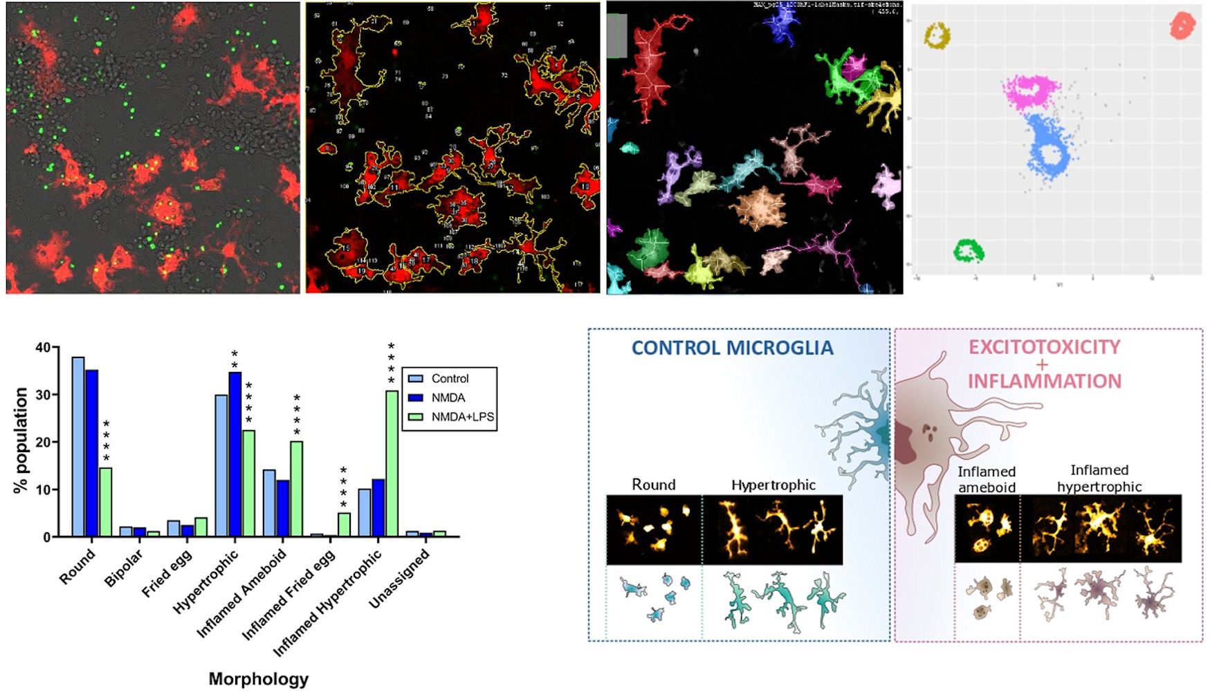
Microglia, the brain's resident immune cell, is highly sensitive to changes in the environment and responds through morphological, functional, and metabolic adaptations. In the brain, microglia coexist with an inflammatory phenotype and with a reparative phenotype, and it is important to be able to detect microglia subtypes in order to design strategies that favor protective forms. In order to quantify the changes that microglia undergo when they go from a healthy environment to a pathological environment, we have developed open access algorithms in ImageJ and R to quantify phagocytosis and identify morphologies.
In this study, we exposed mouse neuron/astrocyte/microglia cell cultures to an excitotoxic, neuron-killing, and inflammatory stimulus and analyzed phagocytosis and morphologies on fluorescence microscopy images after 8h. We stained the DNA fragments released by dead neurons green and quantified their phagocytosis by microglia expressing the red fluorescent protein tdTomato. We identified 7 morphologies: round, hypertrophic, fried egg, bipolar, inflamed amoeboid, inflamed hypertrophic, and inflamed fried egg. We created some programs to separate the different classes and assign a morphology to each microglia in the culture. We observed that in control cultures, round and hypertrophic morphologies predominate. In contrast, excitotoxicity + inflammation causes an increase in inflamed populations and in the proportion of phagocytic microglia. We validate our segmentation and classification programs on mouse brain slices. Our study demonstrates that inflammation is critical to trigger phenotypic changes in microglia. In conclusion, we have generated unique image analysis programs useful in vitro and in vivo that allow discriminating 2 experimental situations through the behavior of microglia.